Analysis was performed on a penicillin G standard and on a penicillin G standard (10 mg/mL aqueous solution) that was decomposed at 60 ºC for 40 hours. The chromatograms are shown in Fig. 1.
The penicillin G peaks virtually disappeared. Peaks 1 and 2 are extremely similar in these mass spectrum patterns. From the MS spectra, the molecular weight is estimated as 308. Because the fragmentation differs, penicillin G and the mother nuclei are assumed not to match.
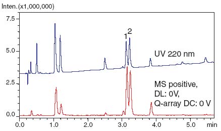
Fig. 1 Chromatogram of Decomposition Products of Penicillin G
MS/MS measurements by LCMS-IT-TOF were performed to predict the structure. Both positive and negative detection indicated C15H20N2O3S as the first candidate.
Benzylpenilloic acids (Fig. 2) are known impurities in penicillin G.
Based on structural predictions, these compounds are thought to be present, as they exhibit similar mass spectral patterns with two adjacent peaks.
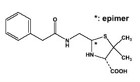
Fig. 2 Structure of Benzylpenilloic Acids